Our Health Library information does not replace the advice of a doctor. Please be advised that this information is made available to assist our patients to learn more about their health. Our providers may not see and/or treat all topics found herein.
Lung Biopsy
A lung biopsy removes a small piece of lung tissue that can be looked at under a microscope. The biopsy can be done in several ways. The method used depends on where the sample will be taken from and your overall health. Methods are:
- Bronchoscopic biopsy. A lighted tool called a bronchoscope is inserted through the mouth or nose and into the airway to remove a lung tissue sample. This method may be used if an infectious disease is suspected, if the abnormal lung tissue is located next to the breathing tubes (bronchi), or before trying more invasive methods, such as an open lung biopsy.
- Needle biopsy. A long needle is inserted through the chest wall to remove a sample of lung tissue. This method is used if the abnormal lung tissue is close to the chest wall. A computed tomography (CT) scan, an ultrasound, or fluoroscopy is usually used to guide the needle to the abnormal tissue.
- Video-assisted thoracoscopic surgery (VATS) biopsy. VATS uses a scope (called a thoracoscope) passed through a small incision in the chest to remove a sample of lung tissue.
- Open biopsy. This method uses surgery to make a cut (incision) between the ribs and remove a sample of lung tissue. It is usually done when the other methods of lung biopsy have not been successful or can't be used, or when a larger piece of lung tissue is needed for a diagnosis.
Why It Is Done
A lung biopsy is done to:
- Diagnose certain lung conditions, such as sarcoidosis or pulmonary fibrosis.
- Diagnose suspected lung cancer. If lung cancer is present, results of the biopsy can determine treatment options (such as surgery, radiation, or chemotherapy).
- Evaluate any abnormalities seen on other tests, such as a chest X-ray or a CT scan. A lung biopsy is usually done when other tests can't identify the cause of lung problems.
How To Prepare
Your doctor may order certain blood tests, such as a complete blood count (CBC) and clotting factors, before your lung biopsy.
Your doctor will tell you how soon before the biopsy to stop eating and drinking. Follow the instructions exactly, or your biopsy may be canceled. If your doctor has instructed you to take your medicines on the day of the biopsy, please do so using only a sip of water.
If you take aspirin or some other blood thinner, ask your doctor if you should stop taking it before your biopsy. Make sure that you understand exactly what your doctor wants you to do. These medicines increase the risk of bleeding.
Be sure you have someone to take you home. Anesthesia and pain medicine will make it unsafe for you to drive or get home on your own.
How It Is Done
A needle or bronchoscopic biopsy can be done without staying in the hospital. You may need to stay in the hospital if you have a video-assisted thoracoscopic surgery (VATS) biopsy or an open biopsy.
You may be asked to remove dentures, eyeglasses or contact lenses, hearing aids, a wig, makeup, and jewelry before the biopsy. You will empty your bladder before the biopsy. You will need to take off all or most of your clothes. (You may be allowed to keep on your underwear if it does not interfere with the biopsy.) You will be given a cloth or paper covering to use during the biopsy. To keep you comfortable, you may get medicines through an intravenous (I.V.) needle in a vein.
A chest X-ray is usually taken after a lung biopsy to look for any problems related to the biopsy.
Bronchoscopic biopsy
A bronchoscopic biopsy is done by a doctor who specializes in lung problems (pulmonologist or a thoracic surgeon). A thin, lighted tool called a bronchoscope is inserted through the mouth or nose and into the airway. The doctor uses the bronchoscope to remove a lung tissue sample.
Needle biopsy
Your doctor will use a CT scan, ultrasound, or fluoroscopy to guide the biopsy needle. The place where your doctor inserts the needle is cleaned first with an antiseptic solution and draped with sterile towels. Your doctor will give you a numbing medicine to keep you from feeling any pain when the needle is inserted into your chest.
Your doctor will then make a small puncture in your skin and ask you to hold your breath while the biopsy needle is inserted into your lung. It is very important to avoid coughing or moving while the needle is in your chest.
After the lung tissue sample is taken, the needle is removed and a bandage is placed over the puncture site. Your care team will position you so that the needle puncture site can seal up. You will need to stay in this position for at least an hour.
Video-assisted thoracoscopic surgery (VATS) biopsy or open biopsy
A VATS or open biopsy is done by a chest (thoracic) surgeon or a general surgeon. You will be kept comfortable and safe by your anesthesia provider. You will be asleep during the surgery.
You will be given a sedative in your I.V. to help you relax about an hour before the biopsy.
An incision is made between the ribs over the area of lung where the tissue sample is to be collected. A scope called a thoracoscope is used in VATS and is passed through this incision to view the surface of the lung and to remove a sample of lung tissue. A larger incision will be made if an open biopsy is needed to remove a tissue sample.
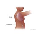
After the tissue sample is collected, your doctor will insert a drainage tube (chest tube) into the area and close the incision with stitches. One end of the tube will be in the space next to your lung. The other end will be sticking out of your chest and connected to a collection container. The chest tube helps re-expand your lung. The chest tubes will be removed when the drainage from your chest has stopped and no air is leaking from your chest incision, usually in a few days. Your stitches will be removed in 7 to 14 days.
VATS uses smaller incisions and takes less time to recover from than an open biopsy.
How long the test takes
Bronchoscopy and a needle biopsy usually take 30 to 60 minutes. You will be in the recovery room 1 to 2 hours.
A video-assisted thoracoscopic surgery (VATS) biopsy or open biopsy takes 1 to 2 hours. Then you will be taken to the recovery room for about an hour. Then you will be taken to a hospital room if you need to stay overnight or longer.
How It Feels
Bronchoscopic biopsy
The numbing medicine used in your mouth or nose generally tastes bitter and may make it hard to swallow.
Needle biopsy
When you are given the medicine to numb your skin at the needle biopsy site, you will feel a sharp stinging or burning that lasts a few seconds. When the biopsy needle is inserted into the chest, you will again feel a sharp pain for a few seconds.
Video-assisted thoracoscopic surgery (VATS) biopsy or open biopsy
If you were given a sedative before the biopsy, you may feel sleepy and relaxed. If you are given general anesthetic, you will be asleep during the biopsy.
Risks
A lung biopsy is generally a safe procedure. Any risk depends on if you have a lung disease and how severe it is. If you already have severe breathing problems, your breathing may be worse for a short time after the biopsy.
Bronchoscopic and needle biopsies are usually safer than video-assisted thoracoscopic surgery (VATS) biopsies or open biopsies. Bronchoscopic and needle biopsies don't need general anesthesia and cause fewer problems. And you don't need to stay overnight in the hospital. But the VATS and open biopsies are more likely to allow a good sample of lung to be removed. A good sample helps determine what the lung problem is and what treatment choices are.
Your doctor will discuss any risks with you.
- Lung biopsy may make you more likely to get a collapsed lung (pneumothorax) during the biopsy. Your doctor may need to place a tube in your chest to keep your lung inflated while the biopsy site heals.
- Severe bleeding (hemorrhage) may occur.
- Spasms of the bronchial tubes can occur, which can cause trouble breathing right after the biopsy.
- An infection such as pneumonia may occur. But usually such infections can be treated with antibiotics.
- Irregular heart rhythms (arrhythmias) can occur.
Results
Lung biopsy results are usually available in 2 to 4 working days. It may take several weeks to get results from tissue samples that are being tested for certain infections, such as tuberculosis.
Normal
- The lung tissue is normal under a microscope. No signs of infection, inflammation, or cancer are present.
Abnormal
- Abnormal cells and tissue in the lung may be present from an active infection, certain lung diseases, or a type of lung cancer.
Related Information
Credits
Current as of: September 29, 2025
Author: Ignite Healthwise, LLC Staff
Clinical Review Board
All Ignite Healthwise, LLC education is reviewed by a team that includes physicians, nurses, advanced practitioners, registered dieticians, and other healthcare professionals.
Current as of: September 29, 2025
Author: Ignite Healthwise, LLC Staff
Clinical Review Board
All Ignite Healthwise, LLC education is reviewed by a team that includes physicians, nurses, advanced practitioners, registered dieticians, and other healthcare professionals.
This information does not replace the advice of a doctor. Ignite Healthwise, LLC disclaims any warranty or liability for your use of this information. Your use of this information means that you agree to the Terms of Use and Privacy Policy. Learn how we develop our content.
To learn more about Ignite Healthwise, LLC, visit webmdignite.com.
© 2024-2026 Ignite Healthwise, LLC.


